Showing posts with label basal. Show all posts

Monday, March 2, 2020

Basal Cell Carcinoma Vs Melanoma

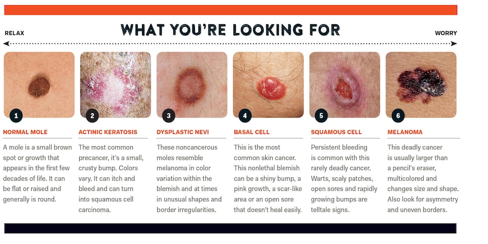
Carcinoma attack basal skin cells bottom level ones and squamous pores and top layer skin cells. The three most common types of skin cancer are in order from most to least.

Skin Cancer Dermatologist In Bethesda Md

This cancer forms in the basal cells one of three types of cells that form the top layer of skin.

Basal cell carcinoma vs melanoma. Melanoma typically begins as a mole and can occur anywhere on the body. Squamous cells which form the outer layer are the other option. Two common skin cancers basal cell carcinoma BCC and squamous cell carcinoma SCC often appear as red patches on the skin similar to rosacea.

The difference between basal cell carcinoma squamous cell carcinoma and melanoma has to do with how the skin cancer cells appear under the microscop. These cells make the brown pigment called melanin which gives. There are multiple types of nonmelanoma skin cancer but the main forms are basal cell carcinoma squamous cell carcinoma and Merkel cell carcinoma.

Major Differences between Melanoma and Carcinoma. Rosacea can both imitate and mask different forms of skin cancer explains Dr. A carcinoma can come from two different types of cell.

This can delay diagnosis which may prove dangerous since early detection of melanoma is critical. Basal cells are the most common culprit. Basal cell carcinoma squamous cell carcinoma and melanoma.

The cells in the bottom layer of the epidermis are the basal cells. Basal cell carcinoma is the most common type of cancer with more than four million cases diagnosed in the United States every year. This type of carcinoma is the rarest form but has the best chance of surviving.

While these make up most of. All three can be caused by the suns ultraviolet rays that damage the skins cells so that they begin to divide uncontrollably. Melanoma is actually a relatively rare form of skin.

They are located in the lower level of the epidermis. What are the treatment options for melanoma. The three most common forms of skin cancer are malignant melanoma squamous cell carcinoma and basal cell carcinoma.

The most important thing about your health is to listen to it constantly if recognized early the survival rates are very high for non-melanoma skin cancer Basal cell and squamous cell carcinoma in the other hand as a second difference there is evidence that the survival rates for melanoma decreases as we get older from 90-95 for 15-39 years old to 80 for men and 85 in women of 70-79 years 4. Basal cell carcinoma is the most common form of skin cancer with approximately 80 of skin cancers developing from basal cells. Over time these lesions may enlarge to become scaly patches of skin that are easily damaged and may bleed frequently.

Tumors overwhelm surrounding tissues by invading their space and taking the oxygen and nutrients they need to survive and function. Basal cell carcinoma appears as a flat pearly bump raised above the skin of the head neck or shoulders. Its main features include redness visible blood vessels and bumps that form mostly on the face.

Neither of those cells can produce a melanoma. 3 The epidermis top layer of the skin has three types of cells. As with nonmelanoma skin cancer exposure to UV radiation from the sun or tanning beds can elevate your risk of developing melanoma but it seems to be less of a factor than with basal cell.

The difference between basal cell carcinoma squamous cell carcinoma and melanoma has to do with how the skin cancer cells appear under the microscop. There are three major types of skin cancers. Carcinoma occurs more frequently in older patients while melanoma more often develops in younger patients.

Carcinomas are identified as either basal cell or squamous cell. As these cells move up in the epidermis they get flatter eventually becoming squamous cells. Amelanotic melanomas can resemble other skin cancers like basal cell or squamous cell carcinoma or worse may be mistaken for benign moles scars or cysts.

Basal cells consistently divide to form new cells. Squamous cell carcinoma may appear as a firm red bump a scaly patch or open sore or a wart that may crust or bleed easily. Their job is to reproduce to create new cells for the skin.

Skin cancers that start in the basal cell layer are called basal cell skin cancers or basal cell carcinomas. Early melanomas are almost always curable while those that advance beyond stage I become more difficult to treat. Basal cell carcinoma may appear as a small white or flesh-colored bump that grows slowly and may bleed.

Like basal cell carcinoma squamous cell carcinoma lesions most frequently develop on parts of the body exposed to sunlight.

Tuesday, July 2, 2019

Basal Cell Cancer Pictures

This picture gallery contains some examples of the more common types of skin cancer as well as some other non-cancerous types of skin growths. But skin cancers can look different from these examples.

Demographics Make A Difference In Common Skin Cancers Medpage Today

A scar-like area that is flat white yellow or waxy in color.

Basal cell cancer pictures. Basal Cell Carcinoma Pictures. It can be pink brown or black. Picture of Basal Cell Carcinoma Basal cell carcinoma.

Superficial or superficiell basal cell cancer looks like a red stain. Below are basal cell carcinoma pictures of typical lesions on various sites of the body. Morphoeic basal cell skin cancer - also known as sclerosing or infiltrating basal cell skin cancer.

You can then show these pictures to your doctor. Skin cancer is the atypical growth of skin cells. This type of cancer typically does not spread to distant sites in the body.

Cancer of the basal cells results in tumors that appear on the skins surface. Time lapse video and pictures of the treatment for my Basal Cell Carcinoma removal skin cancer with no sur. There are different types of basal cell skin cancers.

While BCC almost never spreads to other. Superficial basal cell skin cancer. The cancer may look like a scab or sore that does not heal.

Basal cell skin cancers. Basal Cell Carcinoma Pictures - 54 Photos Images. It can be misinterpreted as a small eczema patch.

It usually on your chest or back but it can also be on your face. These photos and images of basal cell carcinomas are not exhaustive but are examples of common lesions. Pigmented basal cell skin cancer.

The skin appears shiny and taut often with poorly defined borders. This warning sign may indicate an invasive BCC. Micronodular basal cell carcinoma is an aggressive subtype of nodular BCC.

This is why its important to see a doctor if you have any lumps bumps spots sores or other marks on your skin that are new or changing or that. Below are pictures of skin cancer on the neck face and trunk from the left to. Skin cancer close-up - basal cell carcinoma stock pictures royalty-free photos images.

Knot formed basal cell cancer. As the basal cell carcinoma pictures below indicate this type of skin cancer usually shows as a fleshy coloured bump that does not disappear over time and tends to grow slowly in size eventually breaking down and ulcerating. Nodular basal cell skin cancer.

Browse 1072 basal cell carcinoma stock photos and images available or search for skin cancer or squamous cell carcinoma to find more great stock photos and pictures. Basal Cell Carcinoma often look like open sores red patches pink growths shiny bumps or scars and are. It is characterized by multiple small nodules that can be extremely small and uniform with well-defined borders.

More pictures of basal cell carcinoma BCC While the above pictures show you some common ways that BCC can appear on the skin this skin cancer can show up in other ways as the following pictures. A person who has one basal cell carcinoma is at risk for having more. Since not all BCCs have the same appearance these images serve as a general reference to what basal cell carcinoma.

The most common type of skin cancer which commonly presents as a sore that seems to get better and then recurs and may start to bleed. Basal cell carcinoma squamous cell carcinoma melanoma and Merkel cell carcinoma. Micronodular BCC lesions are skin-colored to slightly greyish and may appear a whitish-yellow when stretched.

Basal cell carcinoma is the most common type of skin cancerIt frequently occurs in fair-skinned individuals due to excess sun exposure. Since not all basal cell carcinomas have the same appearance these images should serve as a general reference to what they may look like. Sometimes there can be cuts and scaly like.

Knot formed or nodular basal cell cancer grows deeper into the skin. Basal cell carcinoma may resemble a slowly growing pink skin-colored or light brown nodule on the skin which gradually increases in size. Basal cell carcinoma often occurs on the face and neck where the skin is exposed to sunlight.

At first a basal cell carcinoma comes up like a small pearly bump that looks like a flesh-colored mole or a pimple that doesnt go away. There are four types of skin cancer. The most common cause is prolonged exposure to the sun but it can also develop in areas where no sunlight exposure has occurred.

What does Basal Cell Carcinoma pictures look like. These tumors often look like sores growths bumps scars or red patches. Basal cell carcinoma Basal cell carcinoma skin cancer light micrograph photo under microscope basal cell carcinoma stock pictures royalty-free photos images Pilomatricoma calcified epithelioma neck follicles Pilomatricoma also known as a calcifying epithelioma of Malherbe Malherbe calcifying epithelioma and Pilomatrixoma is a benign skin tumor derived from the hair matrix.

The cancer can be disfiguring if diagnosis and. Basal Cell Carcinoma are abnormal uncontrolled growths or lesions that arise in the skins basal cells which line the deepest layer of the epidermis the outermost layer of the skin.

Lice Bites On Skin

Some people call the critters that gave them this rash sea lice But theyre not lice at all. When a person has body lice on his cloths or a ...